Scattering-Based Imaging
We investigate novel imaging techniques for brain research that exploit the scattering of light to visualize complex brain tissue structures and nerve fiber constellations. Based on simulations with state-of-the-start supercomputers, we develop, build, and test new measurement setups.
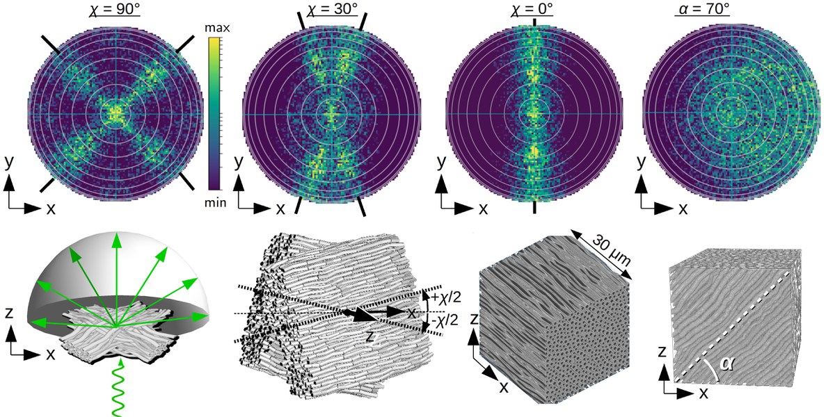
Simulation of light scattering
To analyze the scattering of light on known nerve fiber constellations, we use an electrodynamic simulation approach which models the propagation of light by solving Maxwell’s equations. This allows, for example, to investigate how much the light is attenuated when passing through brain tissue and in which directions the light is scattered. The simulations show that the attenuation of light depends on the direction of polarization relative to the orientation of the nerve fibers – an effect known as diattenuation. Furthermore, the simulations yield characteristic scattering patterns which reveal the orientations of crossing nerve fibers (see figure).
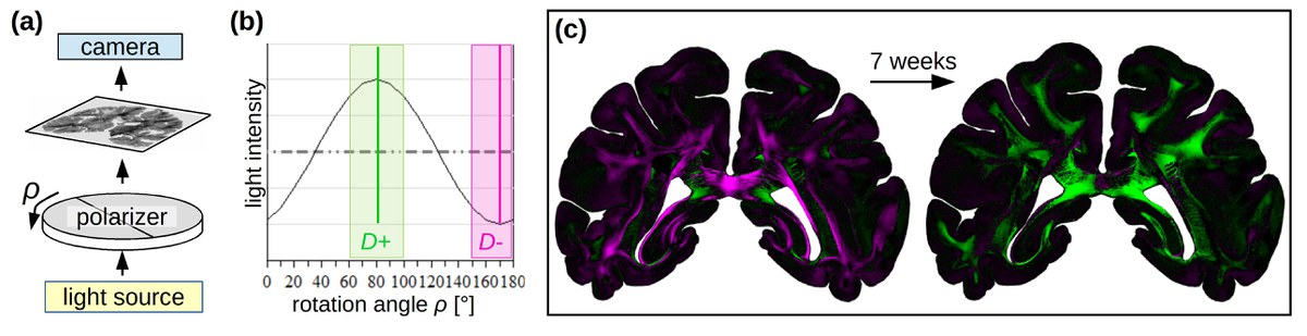
Diattenuation Imaging (DI)
Our imaging technique “Diattenuation Imaging” (DI) makes different tissue types visible. For this purpose, brain sections are illuminated by linearly polarized light (a) and the results are compared to nerve fiber orientations from 3D-PLI measurements: Regions marked in green (magenta) are maximally transparent if the light is polarized parallel (perpendicularly) to the nerve fiber orientation, see figure. How the tissue behaves depends on the time after embedding the brain tissue (c) and also on other tissue properties such as the diameter of the nerve fibers.
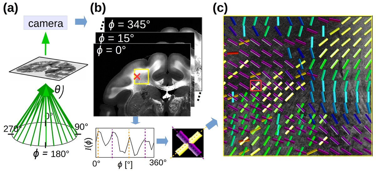
Scattered Light Imaging (SLI)
For our newly developed imaging method “Scattered Light Imaging” (SLI), brain sections are illuminated from different directions and a camera measures the scattered light under normal incidence (a). The positions of the maxima in the resulting intensity profiles (b) provide for each image pixel the directions of (crossing) nerve fibers in the measured brain section (c). To enable a fully automated evaluation of SLI measurements, we have developed a dedicated software (SLIX – Scattered Light Imaging ToolboX).