Deep brain stimulation in Alzheimer’s disease
For Deep Brain Stimulation (DBS) thin electrode rods are stereotactically implanted into defined target regions, mostly in parallel in both brain hemispheres. These are connected to a pulse generator and a permanent high frequency (e.g., 180 Hz) pulsed (e.g., 100 ms width) voltage of e.g., 1.5 V is applied. Depending on location and frequency, neurons of the target region are either inhibited (high frequency) or stimulated (low frequency).
DBS is well established in clinical routine for the treatment of advanced Parkinson’s disease and essential tremor. Particularly, symptoms of tremor, rigor, and bradykinesia are considerably improved. The impressive success of DBS in Parkinson’s disease has prompted to apply this technique in several other clinical indications as for instance obsessive compulsive disorder, Tourette’s syndrome, epilepsy, refractory major depression, chronic pain syndromes and also addictive and eating disorders. Preliminary results in some of these indications suggest that new indications and target structures for DBS are likely to appear in the upcoming years. Interest in DBS is also caused by two key aspects that are clear advantages in comparison to former lesional methods: reversibility and adaptability of stimulation parameters.
However, there are fundamental challenges that actually comprise applicability of DBS: How defining optimal target coordinates and how verifying the therapeutic success in chronic disorders with complex clinical read-out. Experiences from Parkinson’s disease cannot be extrapolated to other disorders, because in Parkinson’s disease correct electrode position and efficient stimulation parameters can be tested during implantation when stimulation effects become immediately visible.
It is therefore necessary to establish surrogate effectors in other disorders to define effective target regions and efficient stimulation parameters. There are molecular and functional imaging methods that have great potentials to help solving these problems. Positron emission tomography (PET) allows measuring metabolic changes in brain tissue caused by DBS including glucose metabolism ([18F]FDG), neurotransmitter receptor function, and perfusion ([15O]H2O).
In a clinical trial, led by the Departments of Stereotactic Surgery and Psychiatry, University of Cologne, DBS of the nucleus basalis Meynert was applied in patients suffering from Alzheimer’s disease. The targeted nucleus is the origin of projection fibres spreading into almost the entire brain and liberating the neurotransmitter acetylcholine. The aim of stimulating the nucleus basalis Meynert was to increase acetylcholine levels and thus to improve brain metabolism and cognitive function.
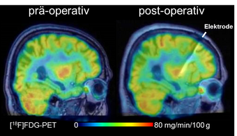
Figure: Superimposed sagittal brain images (MRI, grey-scale and [18F]FDG PET, colour scale) of a patient with Alzheimer’s disease under deep brain stimulation (DBS; left, before surgery; right, after 8 ½ months continuous low-frequency stimulation). The patient (67 yrs.; 3 yrs. since onset of symptoms) showed a clinical improvement that corresponded well with the depicted, remarkably stable, regionally even slightly elevated level of cortical glucose metabolism.
The study revealed that after 12 months DBS several clinical parameters of the patients improved or remained stable. Four of six patients improved clinically, neuropsychologically, and in quality of live. We assessed cerebral glucose metabolism before and 12 months DBS using [18F]FDG and PET and found that responders (clinically improving or stable) showed slight increase of cerebral [18F]FDG uptake in the entire cerebral cortex, namely in the parietal and temporal lobes. This is in contrast to large longitudinal cohort studies that investigated the natural course of Alzheimer’s disease and evidenced an annual decrease of cerebral glucose metabolism of about 5% per year. There was another remarkable finding. Besides obvious long-distance effects on cerebral metabolism, we also observed a local increase of glucose metabolism in the vicinity of active electrode tips contacts that correlated with both cortical effect on metabolism and clinical response. This finding corroborates the concept of low frequency stimulation of grey matter areas resulting in cellular activation.