Atomic Force Microscopy
Atomic force microscopy is a technique used to mechanically scan surfaces on the nanometer to micrometer scale and measure atomic and molecular forces.
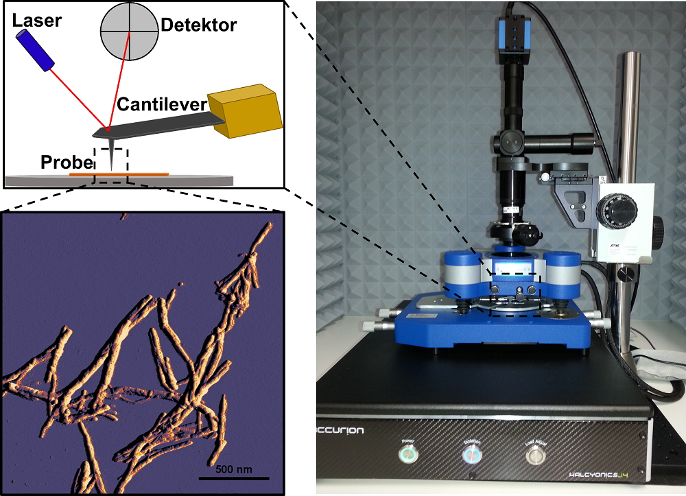
An atomic force microscope scans the texture of a surface with a nonoscopically small needle (see Fig. above left). This needle, attached to a leaf spring (cantilever), is moved over the surface by piezo crystals. The leaf spring bends at the surface structures. This bending of the leaf spring (deflection) is measured by a laser, which is focused on the tip of the leaf spring, and optical sensors. This produces a surface profile.
Imaging of surfaces can be operated by means of different modes. In contact mode, the AFM tip is in direct contact with the electron shells of the atoms on the surface. In intermittent mode (tapping mode), the AFM tip is excited close to the resonance frequency. The AFM tip, now oscillating continuously at a defined amplitude, scans the surface. Both intermittent and contact modes can be applied to air-dried surfaces or surfaces in buffer. Especially the latter option is a suitable method to image biological samples, even living cells, in native or near-native environments on a nanometer scale.
If the AFM tip is pressed onto a point on the surfaces with a defined force and then lifted again, force-distance curves can be recorded in this way. The dependence of the force acting on the AFM tip on the tip position recorded in this way allows conclusions to be drawn about the material properties of the samples, such as adhesion or elasticity. Applying the method of force-distance curves to single molecules provides information about the binding forces in single molecules. In this way it is possible to study e.g. protein folding or protein unfolding.